The Central Canal
The central canal runs through the spinal cord and is filled with CSF, effectively transporting nutrients throughout the spinal cord. It is connected to the ventricular system, and is lined with ependymal radial glial cells (ERGs) and CSF contacting neurons (CSF-cNs). The ERGs apical extension bears a motile cilium, which is believed to be responsible for the motion of CSF inside the canal. CSF-cNs also posses a cilium, believed to monitor certain parameters of the CSF, such as acidity, heat, bacterial metabolites and spinal curvature. The following animation represents a 1 dpf larvae, with the CC and the ventricular system highlighted in green, and CSF-cNs in purple.
CSF Flow in the Central Canal
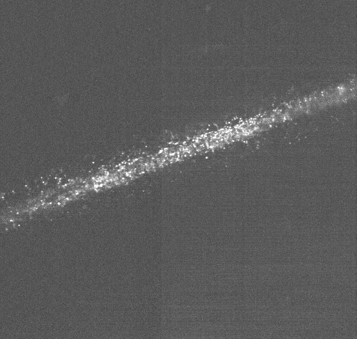
CSF in the central canal has a bilateral flow profile. It flows towards the tail ventrally, and towards the head dorsally. As mentioned previously, this particular flow profile is believed to be generated by ERGs motile cilium. Below is a video taken in vivo of a 1 dpf wild type larvae injected with fluorescent nanobeads.
What is a kymograph?
For quantifying how CSF flows in the central canal, we want to track the injected particles in a 2D plane over a certain amount of time. To acheive this we first need to acquire a time lapse of the central canal. Next, we could use classical particle tracking algorithms to track individual beads and infer their velocity. But given the high amount of noise in our images, these methods have proved to be poorly efficient. Instead, we use kymographs.
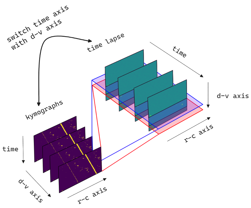
By re-arranging the pixels of our time lapse in a certain manner, we are able to visualize the trajectory a particle/bead takes in one plane over time. This gives us a position as a function of time graph, from which we can extract the direction and velocity of our particles.
To generate a kymograph, the time axis of our time lapse is exchanged with the dorsal-ventral axis. This gives us an array of kymographs, that represents the trajectory of a particle in the rostro-caudal plane over time for every dorso-ventral position. For each of these kymographs we perform blob detection and from each blob's orientation angle, we can extract the particle's velocity. Although we only detect velocity along one axis, and one same bead may be detected multiple times, this method still gives us valuable insight on CSF flow dynamics. This whole analysis process was implemented in python and the code can be found here.